

Phone:
0086 28 87573647
+86 19150187139
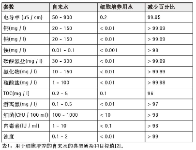
There are many sources of contamination in water used for cell culture, such as bacteria, yeast, or mold. These contaminants are usually visible to the naked eye or under an optical microscope. However, chemically derived contaminants or other biological agents can also affect the growth, morphology, or cell behavior of cultured cells, but they are not detectable to the naked eye. Therefore, water used for cell culture must be free of microorganisms, especially endotoxins, inorganic ions (heavy metals such as lead and zinc), or organic compounds (humic acid, tannic acid, pesticides, etc.). For more information, please refer to references [1, 2]. Typical impurities in tap water and target values for cell culture are shown in Table 1.
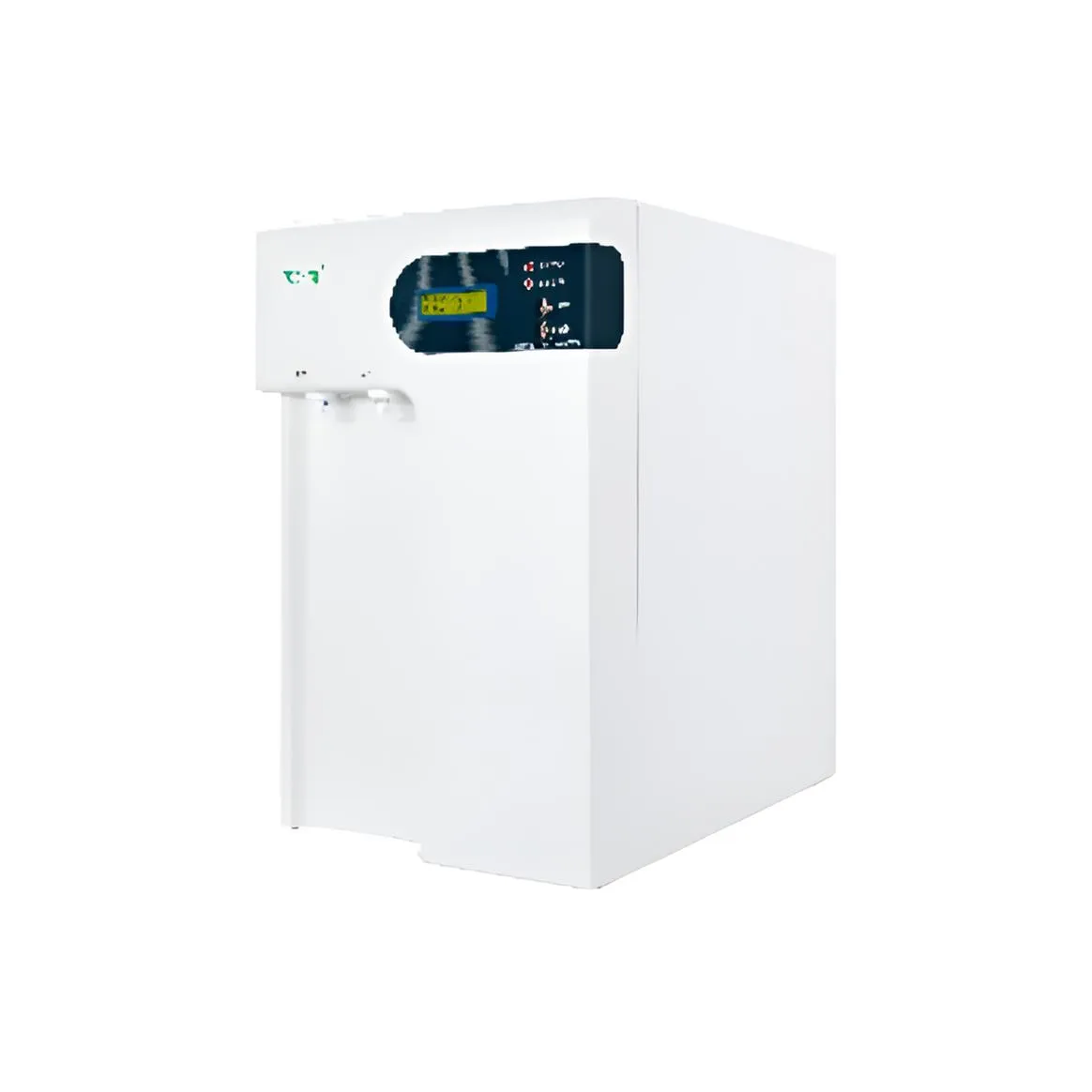
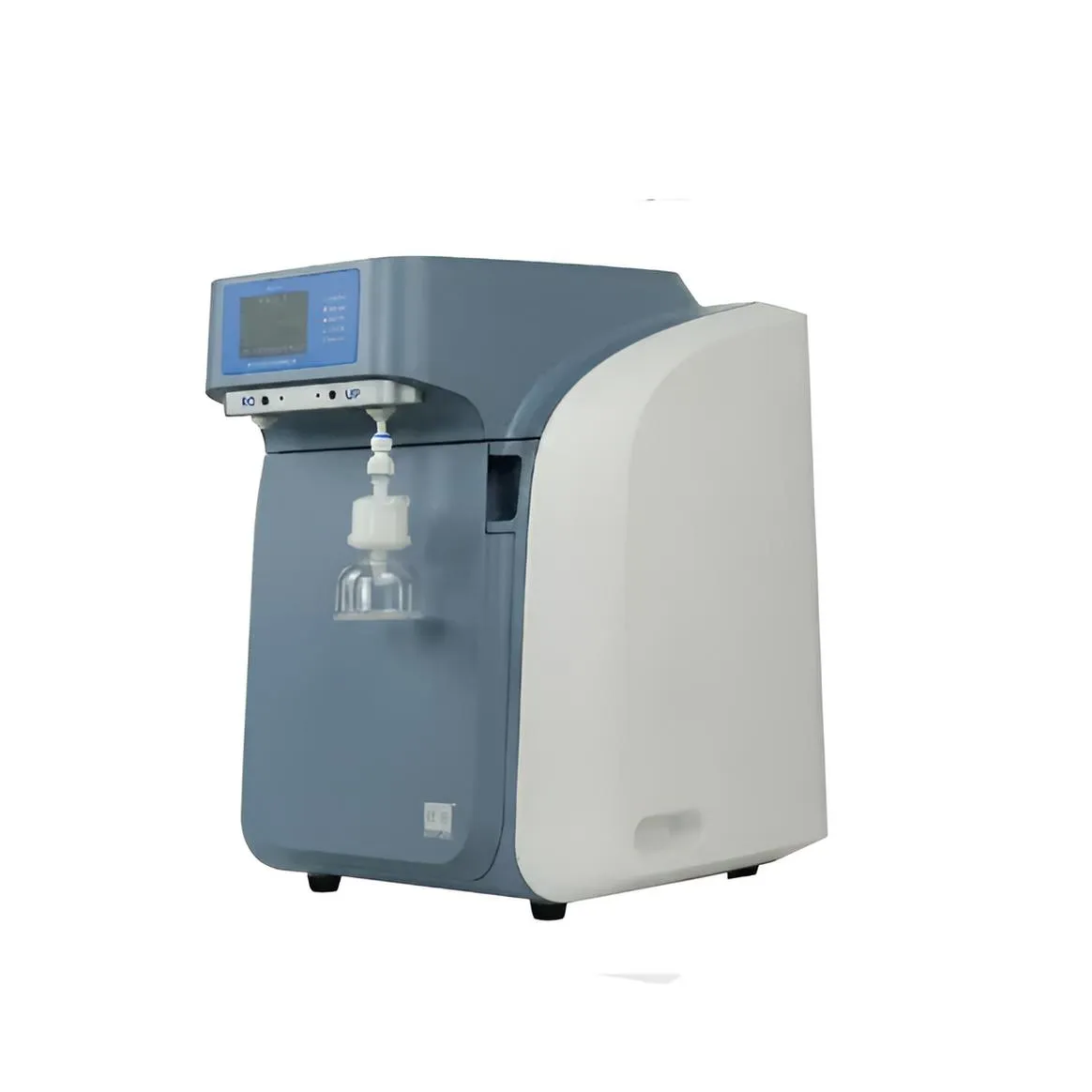
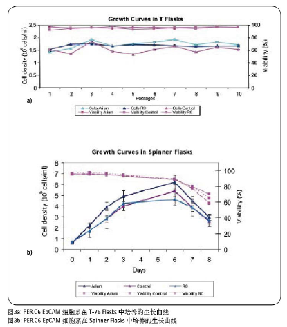
The purpose of this experiment was to evaluate whether the ultrapure water produced by the AriumproUF system can be safely and easily used for cell culture applications. In this study, we used ready-to-use CDM4PERMab (Hyclone) medium to culture PER.C6EpCAM cells as a control, and prepared CDM4PERMab (Hyclone) powder medium using water produced by AriumproUF (UF water) and RO water, respectively. The RO water data used in this application guide were obtained using the older model of the next-generation Arium Advanced system (AriumRO). The results of each culture batch were used to evaluate whether the water produced by the AriumproUF system was suitable for PER.C6EpCAM cell culture.
The PER.C6 cell line used in this experiment is derived from human retinal cells and is now also used for recombinant protein expression and monoclonal antibody (Mab production) preparation, as well as for the production of therapeutic proteins and monoclonal antibodies.
Conclusion
The results of this experiment clearly demonstrate that the powdered culture medium (CDM4 PERMab medium) prepared with UF water is more suitable for culturing the PER.C6EpCAM cell line than commercially available ready-to-use media. The growth characteristics of the PER.C6EpCAM cell line cultured in UF water medium were similar to those of the control group cultured in ready-to-use CDM4 PERMab medium. Furthermore, we observed that when the experiment was conducted in SpinnerFlasks, cell samples cultured in UF water medium showed better cell growth compared to those cultured in RO water medium. In this experiment, cell density was generally very high, and O2 was no longer a limiting factor. Therefore, we conclude that RO water, due to its higher concentration of endotoxins or inorganic salts, affects cell growth.
These experimental results are further validated and demonstrated by the monoclonal antibody yield obtained by culturing the PER.C6EpCAM cell line in SpinnerFlasks. The PER.C6EpCAM cell line achieved the highest monoclonal antibody yield when cultured in a medium prepared with AriumproUF ultrapure water, while the yield in the control group (using ready-to-use medium) was slightly lower. The monoclonal antibody yield in both cases differed from that in the RO water sample; in fact, the monoclonal antibody yield decreased after culturing in the RO water sample.
In summary, we conclude that the AriumproUF system is the most suitable for cell culture of the PER.C6EpCAM cell line because this water system minimizes the content of impurities in the water, such as inorganic ions and organic compounds, especially reducing endotoxin levels to extremely low levels. This result has also been verified in the latest experiments [4].
ULPHW Ultra-Low TOC Series Ultrapure Water System – Your “Big Gift” Event is in Full Swing!
References/More Information
[1] ASTM Standard Guide for Bio-Applications Grade Water D 5196-06
[2] Whitehead P.: Water Purity and Regulations, in Medicines from Animal Cell Culture (eds. Stacey, G. and Davis, J.), John Wiley & Sons, Ltd (2007)
[3] Freshney I. R.: Culture of Animal Cells – A Manual of Basic Technique and Specialized Applications-6th edition, John Wiley & Sons, Inc., USA (2010)
[4] Schmidt K. und Herbig E.: “Weniger ist mehr –Quantitative Endotoxinbestimmung von Reinstwasser“, Laborpraxis, 5, 36. Jhg., (2012)